


Actinic Keratosis (Sun Damage) – Examination and Treatment by a Dermatologist
A specialist in dermatology examines rough, scaly areas of sun-damaged skin and treats them with freezing, curettage or topical cream.
Actinic keratosis (solar keratosis) consists of rough, scaly patches of skin caused by many years of sun exposure. The patches are typically found on the face, ears, a bald scalp and the backs of the hands. Actinic keratosis is a precancerous skin change that can, in rare cases, develop into squamous cell carcinoma of the skin. At Charlottenlund Privathospital, actinic keratosis is examined by Susanne Birkballe, specialist in dermatology, using a dermatoscope and, when needed, a tissue sample. Treatment may include freezing, curettage or topical cream, with follow-up arranged as needed.
What is actinic keratosis?
Actinic keratosis develops when ultraviolet radiation from the sun damages cells in the outermost layer of the skin over many years. The cells begin to divide abnormally, resulting in a rough, scaly patch. The abnormality is confined to the epidermis and is not cancer, but it is regarded as a precancerous change that can develop into squamous cell carcinoma of the skin.
According to the European guideline, actinic keratosis is the most common new skin growth in people with fair skin. The patches often occur within areas of skin that are generally sun damaged. For this reason, the dermatologist looks not only at the individual patch but at the entire sun-exposed area.
Source: Flohil et al., Rotterdam Study, Journal of Investigative Dermatology 2013
Who gets actinic keratosis?
Actinic keratosis is seen particularly in people over 50 with fair skin that burns easily. The risk increases with cumulative sun exposure over a lifetime, including outdoor work, sport, holidays in sunnier countries and use of sunbeds. Men develop actinic keratosis more often than women, and a bald scalp is particularly exposed.
People with a weakened immune system, for example after an organ transplant, have a higher risk of both actinic keratosis and skin cancer and should have their skin examined regularly.
What does actinic keratosis look like?
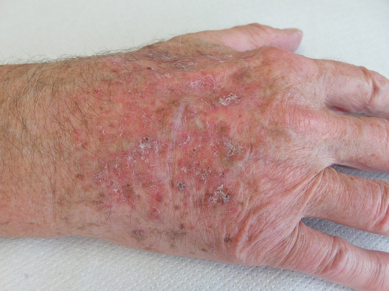
Actinic keratosis appears as a rough, scaly patch that may be red, skin-coloured or brownish. It is typically a few millimetres to a couple of centimetres in size. The patch may be easier to feel than to see because it can feel like sandpaper when you run a finger over it. Some patches may sting or itch slightly.
The patches occur on skin that has received a lot of sun exposure: the face, nose, ears and forehead, a bald scalp, the backs of the hands and the forearms. When the lower lip is affected, the condition is called actinic cheilitis, which causes dry, scaly and pale skin.

Grade 1 – thin spot
Easier to feel than see
Mild redness and fine scales
Grade 2 – visible stain
Clearly visible and easy to feel
Rough, scaly surface
Grade 3 – thick spot
Hard and thickened
Thick scales; may look like a small horn
Actinic keratosis can be confused with other skin conditions, such as age spots (seborrhoeic keratoses), eczema, psoriasis or rosacea. A dermatoscope examination can distinguish between them.
When can actinic keratosis develop into skin cancer?
Most spots with actinic keratosis never develop into cancer, and some disappear on their own. But a small number can develop into squamous cell carcinoma of the skin, and it is not possible to tell in advance which spots will do so.
In a US study of 169 patients with a total of more than 7,000 spots on the face and ears, the risk of an individual spot developing into squamous cell carcinoma was 0.6% after one year and 2.6% after four years. However, around 65% of the squamous cell carcinomas that developed came from a spot previously diagnosed as actinic keratosis.
For this reason, European guidelines generally recommend treatment, and spots that change should be checked.
When should you see a dermatologist?
You should see a dermatologist if you have rough, scaly spots on sun-exposed skin that do not go away, or if you have many of them. A spot should always be checked if it gets thicker, grows quickly, becomes tender, bleeds or turns into a sore that does not heal. This also applies to spots that come back after treatment and new bumps on the skin that you are unsure about—including moles that change.
If you have had skin cancer before or have a weakened immune system, it is a good idea to have your skin checked regularly.

How does a dermatologist examine actinic keratosis?
The examination begins with a conversation about sun habits, previous skin cancer and how long the spots have been there. The skin is then examined—not only the spots you have noticed yourself, but also the rest of the sun-exposed skin.
The spots are examined with a dermatoscope, a magnifying glass with a light that reveals patterns in the skin that cannot be seen with the naked eye. If a spot looks different, is very thick or has not gone away after treatment, a small tissue sample is taken under local anaesthetic. Results are usually available within 5–10 working days.
The examination is carried out by Susanne Birkballe , a dermatologist. If the tissue sample shows skin cancer, further treatment will be planned.
How is actinic keratosis treated?
Treatment is chosen based on how many spots there are, how thick they are and where they are located. Individual spots are often treated by freezing them with a very cold liquid. A small blister or scab may form and heal within a few weeks. Thick spots can be scraped off so the treatment reaches deeper.
When there are many spots in the same area, such as on the forehead or scalp, cream treatment is often used, with the cream applied to the whole area for a period of time. The cream also treats changes that are not yet visible. The skin typically becomes red and irritated during treatment, and the dermatologist will explain what to expect.
Follow-up appointments are arranged as needed to assess the effect of treatment and check the skin for new spots. Rough bumps on the hands may also be warts, which are treated differently.
How can actinic keratosis be prevented?
The most important prevention is to protect your skin from the sun: seek shade during the middle of the day, wear a hat with a brim and covering clothing, and apply sunscreen to your face, ears, hands and bald scalp. Avoid tanning beds. It is never too late to start, and good sun protection can reduce the number of new spots.
It is also a good idea to check your skin regularly and look out for new spots or spots that change.
Referrals and payment
Many health insurance policies cover a dermatologist’s examination, but insurers typically require a referral from your GP and prior authorisation. No referral is needed if you are paying privately. Prices are listed in the price list. Consultations take place at the main clinic in Charlottenlund.
Frequently asked questions
Is actinic keratosis dangerous?
Actinic keratosis is not cancer, but a precursor that can in rare cases develop into squamous cell carcinoma of the skin. Treatment of the spots is therefore generally recommended, and you should have your skin checked if a spot changes.
What does actinic keratosis look like?
Typically, it appears as a rough, scaly spot that is red, skin-coloured or brownish. It is found on skin that has had a lot of sun exposure, such as the face, ears, bald scalp and backs of the hands. The spot can often be felt more easily than seen, as it feels like sandpaper.
How does a dermatologist treat actinic keratosis?
Individual spots are frozen or scraped off. When there are many spots in the same area, the entire area is often treated with cream. The dermatologist chooses the treatment based on the number, thickness and location of the spots.
What grades of actinic keratosis are there?
Actinic keratosis is divided into three grades according to thickness: grade 1 spots are thin and easier to feel than see, grade 2 spots are clearly visible and rough, and grade 3 spots are thick, hard and scaly.
Can actinic keratosis come back?
Yes. The skin around the spots is often sun-damaged too, so new spots may appear. Follow-up appointments are arranged as needed, and good sun protection is important.
When should a spot be checked for skin cancer?
A spot should be checked if it gets thicker, grows quickly, becomes tender, bleeds or turns into a sore that does not heal. This also applies to spots that do not go away after treatment.
Read also
Other skin changes examined by the dermatologist:
→ Dermatologist and dermatology – Overview of skin conditions → Moles – Assessment and removal → Warts – Rough bumps on the hands and feet → Rosacea – Redness and spots on the face